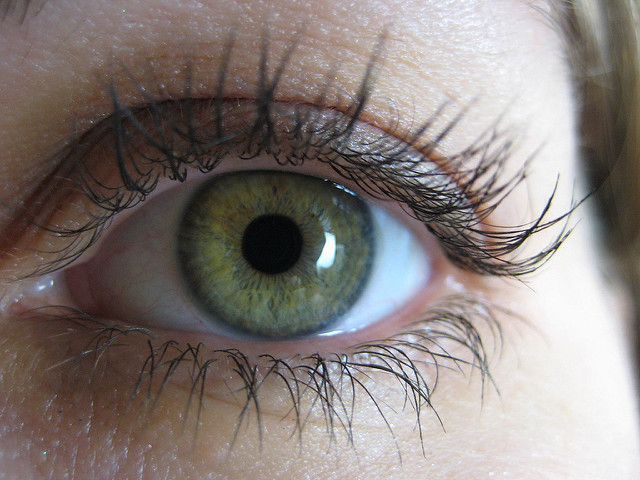
Our eyes are one of the most complex organs in the body. Our vision relies primarily on our eyes’ ability to detect light using the retina, a thin tissue layer located at the back of our eyes. In patients with lattice degeneration, this tissue begins weakening at the outer edges, and leads to tears in the retina, called atrophic holes. This weakening forms in the pattern of a lattice, giving the disease its name. Normally, we only use the central portion of the retina, but the danger in lattice degeneration lies in the fact that it can increase the risk of retinal detachment, which causes permanent blindness.
Unfortunately, there are no known causes of lattice degeneration. However, myopic (near-sighted) patients are often diagnosed with this disease, potentially as a result of the unusual shape of their eyes. In addition, it seems to be passed down through families. There are also no symptoms associated directly with lattice degeneration. Instead, symptoms only appear when the disease progresses to a more serious state, involving retinal detachment.
Image Source: Callista Images
A retinal detachment can be caused when the clear gel, or vitreous humor, that normally fills the eye, seeps into atrophic holes and causes detachment of the retina. Patients should look out for common symptoms that can indicate a retinal detachment:
- Blurred vision
- Floaters: small dots or shadows that float around in your field of vision
- Loss of peripheral vision
- A significant overall decrease in vision
There is no cure for lattice degeneration, but serious atrophic holes can be treated with either cryotherapy or photocoagulation. Cryotherapy involves freezing the tear with a cold probe. Photocoagulation is a laser surgery that uses a highly focused beam of light. During these procedures, the surgeon seals off the hole in the retina by forcing localized scarring in the tissue around the hole. The scarring acts like a fence to prevent the vitreous gel from entering and pooling in the holes. Since the surgery does not prevent further holes from appearing, patients should continue to be aware of the symptoms of retinal detachment.
As a patient with lattice degeneration, I always worry about the imminent risks of retinal detachment. It’s frightening how easily we can lose our vision. Therefore, patients with lattice degeneration should check with a retinal specialist once a year to watch for any new atrophic holes and to monitor the size and position of previous atrophic holes. And, even if you don’t have lattice degeneration, please be aware of the symptoms and be sure to attend your annual medical checkups! Your eyes will thank you for your foresight!
Feature Image Source: Eye by madaise


