


Is it possible to enter a dream state whenever we’d like?
Neuroscientists at UC Berkeley can send a sleeping mouse into the land of dreams. Their study showed that an optogenetic switch (on/off switch for neurons) implanted into nerve cells of the brain’s medulla can activate or inactivate neurons. With neuronal activation, the sleeping mice entered REM sleep within seconds. REM sleep is a dream state in mammals characterized by rapid eye movements, cortex activation, and complete paralysis of the skeletal muscles. Inactivation of these neurons in the medulla barred the ability to enter REM sleep altogether, or minimized it.
Image Source: H. Armstrong Roberts.
In fact, author Professor Yang Dan added that 94% of the mice entered REM sleep in seconds, concluding that a rather small network of neurons made the decision to dream.
Researchers believe that this finding will help us understand the dream-sleep cycle, and uncover the secrets of “why we dream”. Many mental disorders are linked to REM sleep, and numerous drugs affect it. Postdoctoral fellow Franz Weber hopes the study can be used to find cures to neurological diseases like Parkinson’s and Alzheimer’s.
Activating the neurons during wakefulness did not affect the mice, except for increasing their appetites. In normal mice, these neurons release the neurotransmitter GABA. GABAergic neurons are most active when mice are eating or grooming, two highly pleasurable activities. Professor Dan suspects that GABAergic neurons in the medulla have the contrary effect of stress neurons, like the noradrenergic neurons in the pons releasing noradrenaline. She adds that noradrenergic neurons are active during activities like running, and shut down during activities like eating and grooming. During relaxation and enjoyment, the noradrenergic neurons switch off, and GABAergic neurons turn on.
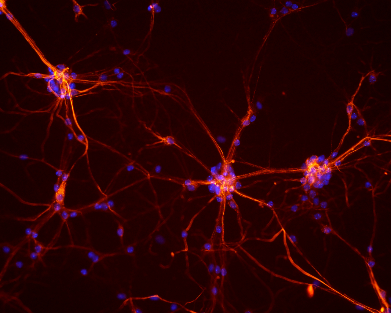
Through optogenetics, genetically engineered mice with a marker protein in specific neurons were used to target a virus to the GABAergic neurons. By shining a laser light through an optical fiber inserted in the brain, an ion channel can activate the neurons; alternatively, neurons can be deactivated by inserting an inhibitory ion pump into the GABAergic neurons. This has helped to map brain activity and study sleep-wake behavior.
Image Source: Science Photo Library – KTSDESIGN.
Using drugs rather than optogenetics to activate the same neurons reduced REM sleep as well, but the drugs were slower to take effect, and wore off. Inserting light-sensitive ion channels into glutamatergic neurons, which release glutamate, immediately awakened the mice; this is the opposite effect of activating GABAergic neurons. The paper is published in the journal Nature, and Professor Dan’s team is currently continuing to study the neurons that affect REM and non-REM sleep.
Featured Image Source: NICHD