Our eyes are complex structures that transform light into meaningful electrical signals that our brains interpret as images. Incoming light passes through the eye until it hits a layer of photosensitive cells, called the retina, along the back wall of the eye. The rods and cones in the retina are photoreceptors that are responsible for processing light. Rods are more numerous than cones and provide vision in low-light conditions, but they don’t help with color vision. Cones provide color vision and are split into three subgroups that process different wavelengths of light: red, blue, and green. Rods are mostly used for nighttime vision because they require less light than cones.
Image Source: Steve Gschmeissner
An intuitive design would place the rods and cones in the front layer of the retina where they can be exposed to as much light as possible and the image-processing neurons to be behind them. However, the rods and cones in most vertebrates are in fact in the very back layer of the retina, covered by dense layers of neurons and cell bodies. Their prevalence suggests an evolutionary advantage to having the photoreceptors stashed in the back, away from light.
A study done by Dr. Erez Ribak and Amichai Labin from the Technion, Israel Institute of Technology investigated this “backwards wiring” by comparing how light is processed in the retina between humans and guinea pigs, which are also active during the day and have well-characterized retinas. They built a computer model of the human retina and saw that a layer of Muller glial cells, cells that support the surrounding neurons, act as fiber optic cables that pipe the light to the rods and cones in the back of the retina. “Further computer simulations showed that green and red are concentrated five to ten times more by the glial cells, and into their respective cones, than blue light. Instead, excess blue light gets scattered to the surrounding rods,” Ribak writes. They tested their findings by passing light through the guinea pigs’ retinas and examining the results through a microscope in three dimensions. The results confirmed an uneven distribution of light in the retina, with red and green light being concentrated in columns of light through retinal layers up to ten times the average intensity. Blue light, which was less readily transmitted through the glial cells, was scattered among the surrounding rods.
These findings suggest that the structure of the eye has been optimized to increase the concentration of cells with the right length and density to intensify specific wavelengths of light, enhancing daytime vision without hurting nighttime vision. However, Dr. Mark Hankins, a professor of visual neuroscience at Oxford, is not entirely convinced that Dr. Ribak’s team’s findings are the “driving-force” behind the “backwards wiring” of the eyes. He points out that the non-intuitive structure can also be attributed to increased efficiency in the disposal of worn-out cell components and the photoreceptors’ need for a large source of energy. According to BBC, Dr. Hankins goes on to say, “We should also remember that several animal classes do not have a ‘backward-pointing’ eye, and also have Muller cells” (Jonathan Webb, BBC).
The research paper can be found here.
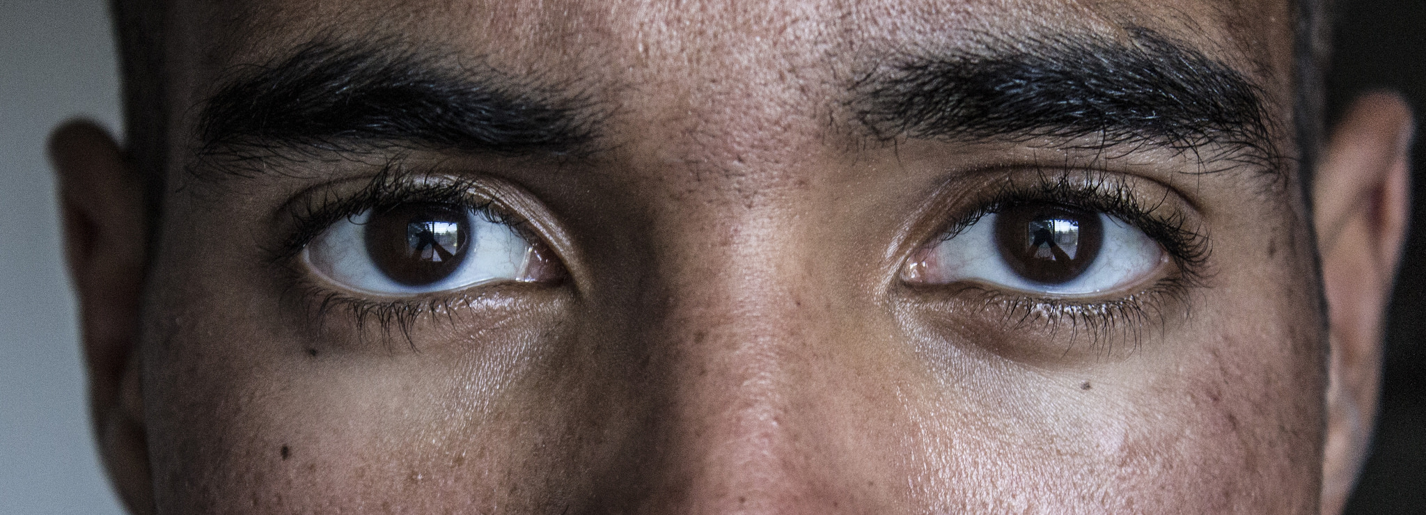
Feature Image Source: Eyes by Dboybaker


