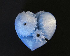
Amidst all of the buzz of the newly developing 3D printing technology comes a new cutting edge idea: 3D printed hearts. For the 40,000 babies born each year with congenital heart defects (CHD), or defects in the heart that are present at birth, 3D printed hearts are a new and innovative option that can be utilized to help save their lives.
Image Source: Morsa Pictures
Before performing surgeries, doctors use computer models to visualize the heart in order to develop a course of action for the surgical procedure. Experts on congenital heart defects at Spectrum Health Helen DeVos Children’s Hospital have developed a new hybrid 3D model of a patient’s heart by combining computed tomography (CT) scans and three-dimensional transesophageal echocardiography (3DTEE) scans. CT scans use a combination of x-rays taken from different angles to form images of cross-sections of bones, blood vessels, and soft tissues in the body. 3DTEE scans provide a 3D visual of the patient’s heart anatomy. The CT scans provide the best visualization of the anatomy of the outside of the heart, whereas the 3DTEE scans provide a superior visualization of the anatomy of the heart valves. Combined together, they can be used to render a 3D printable model of a CHD patient’s heart. Using this heart, surgeons can then create a more accurate and developed procedure to conduct the surgery needed to repair the congenital heart defects.
Proof of concept studies have also brought up the possibility of combining this hybrid with a third scanning method: magnetic resonance imaging (MRI). MRI uses magnetic pulses and radio waves to render 3D images of organs and structures in the body. It’s currently the best imaging technique for the inside of the heart. When the MRI is combined with the CT and 3DTEE scans, the hybrid 3D model of the patient’s heart produced truly holds the potential to produce a superior 3D printed heart model for surgeons looking to operate on the heart.
Feature Image Source: 3D Printed Heart Gears by John Abella


