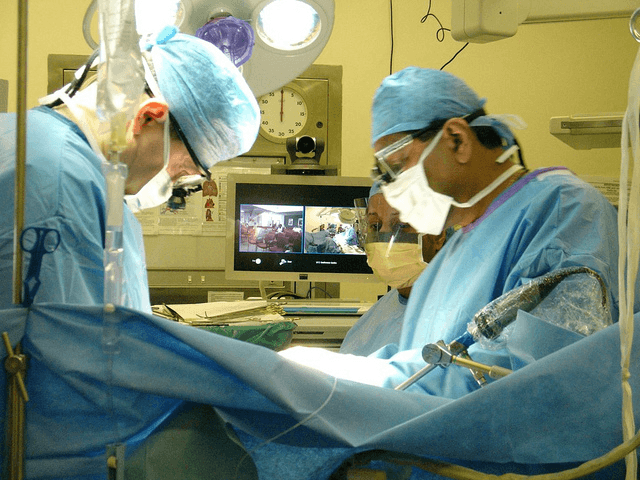
An endoscope is a medical tool used to internally examine a patient’s body. It’s a thin, hollow tube that has a small camera and light attached to one end. The tube is inserted into the patient’s body and the image is fed back to a screen from the camera. Doctor’s can use the endoscope to confirm a diagnosis, conduct a biopsy, or even perform surgery. Soon, however, the tool may have a new purpose.
Image Source: Isa Foltin
Researchers at the University at Buffalo are developing a new endoscope that can provide better images as well as the ability to assist in attacking tumors. The new imaging technology projects patterns of different frequencies of light on the cell. These improved imaging techniques result in a high-contrast picture of the tumor.
In order for the endoscope to assist in destroying tumors, chemotherapy drugs are first delivered to the body intravenously, or through the veins. However, the drugs will be contained in nanoballoons, capsules that open upon light exposure. This way, the side effects of chemotherapy will be reduced as the encapsulated drugs will not affect healthy cells in the body. Once the nanoballoons reach the tumor, doctors can use the endoscope to shine a beam of light on the capsules, allowing them to open and release the chemotherapy drugs onto the tumor cells.
Dr. Ulas Sunnar, the principal investigator on this project, is currently working on making it easier for doctor’s to control the light from the endoscope. They are creating a “digital mask” which will be able to adjust the intensity and shape of the light beam.
The mask is sort of like the Bat signal from Batman movies. It alters the shape of the light. At the same time, we’ll be able to control the strength of the light. The combination will allow us to manipulate the beam to target cancer cells with unprecedented accuracy.
-Dr. Ulas Sunnar
Feature Image Source: DSC06002 by Andy G


