


The use of human stem cells has been revolutionizing the way in which scientists are able to study the human body and diseases. Researchers can use stem cells in order to develop organoids, three-dimensional organs grown in a petri dish (in vitro), that can be used to model human organs. Recently, a research team from Harvard University was able to use both human embryonic stem cells (hESC) and human induced pluripotent stem cells (iPSC) in order to develop blood vessel organoids that can express a diabetic phenotype, or a physical/viewable characteristic of the disease.
Diabetes is a common disorder in which people either are unable produce insulin (type 1 diabetes) or their body has grown insensitive to the insulin that is released by the pancreas (type 2 diabetes). As a result, elevated levels of sugar are found in the bloodstream, so patients have to inject insulin in order to control their blood sugar levels. This research group first established a method for growing a blood vessel organoid from stem cells in a non-diabetic environment. These vessels were then transplanted into mice, where they were able to further grow and develop into more advanced vascular structures like capillaries and arteries.
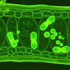
Image Source: Sitox
Next, the research team introduced the cultured vessel organoids to an environment that would mirror the elevated blood sugar levels found in a patient with diabetes. In response to the elevated sugar levels or “diabetic environment”, the cultured vessel organoids phenotypically changed and the blood vessel membranes increased their thickness. This is a very common phenomenon in diabetic patients and can lead to many complications due to the lack of oxygen diffusing to tissues; thicker blood vessels impair the travel of gases from the vessels into tissues. The phenomenon can eventually lead to amputations of the lower portion of the leg as the tissue becomes necrotic, or starts dying, from the lack of oxygen and nutrients being delivered. The team then used widely prescribed drugs for diabetes to see if they could reduce the vessel thickening. There were no promising results found with the anti-diabetic drugs, but the investigation found a protein called gamma-secretase that appeared to be involved in the process of vessel thickening.
This research is a revolutionary step forward in the ability to study vascular diseases and model a human organ in a petri dish. The ability to model an organ then allows for the ability to have a deeper understanding of the molecular mechanisms that lead to the expression of distinct phenotypes involved with disease, which can then translate over to the development of further therapies and treatments.
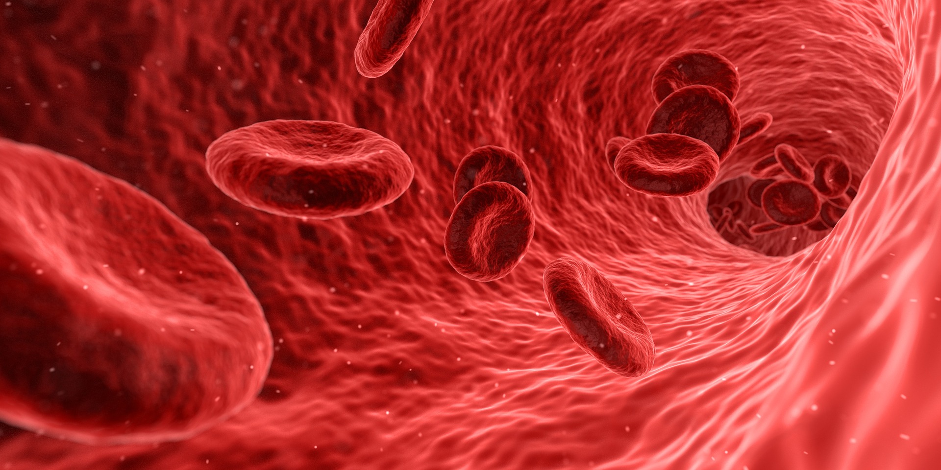
Feature Image Source: Arek Socha