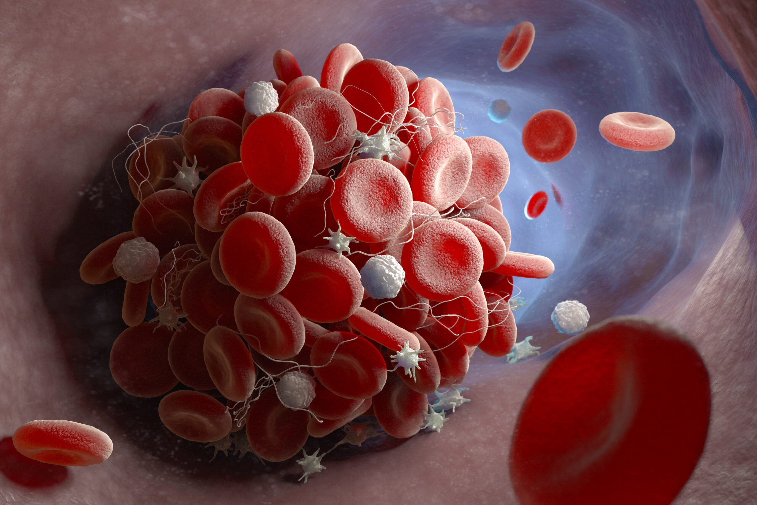
Atrial fibrillation is a condition that can be rather tricky to diagnose as it can be difficult to detect the irregular heart beating in a clinical setting, oftentimes being misdiagnosed or undetected. This is further complicated by the fact that atrial fibrillation is often asymptomatic, which further adds to this condition being missed by physicians and undiagnosed. The main concern with atrial fibrillation is that the irregular beating can promote blood clot formation in the heart, and beating can then dislodge the clot, increasing the risk of a stroke. A team from Massachusetts General Hospital has successfully developed a new imaging tool that aids in the early detection of blood clots.
With regards to blood clot detection, the standard procedure can be a rather invasive process for patients to undergo as a scope has to be inserted orally which requires patients to undergo anesthesia. This new technique is classified as a contrasting agent as it can be injected into the bloodstream and then subsequently imaged, creating a noninvasive procedure for patients. The tag has a strong binding affinity to a protein found in blood clots called fibrin, which is a protein that helps bind blood cells together to form a clot. The radioactive copper probe can then be imaged using a positron emission tomography (PET) scan, which can enable blood clot detection not only in the heart but also in the entire body. The tag is then naturally degraded by the body after a couple of hours, making this procedure safe for patients.
A PET scan is commonly used to detect the metabolic activity in the brain and heart, and now can be used to detect blood clots of this new bioimaging tag.
Image Source: JohnnyGreig
Working to develop new techniques like this helps increase the power of preventative medicine. Detecting blood clots before they migrate to other places in the body can be a life-saving procedure, specifically by preventing a stroke from atrial fibrillation complications. Furthermore, creating less invasive measures can increase patient comfort and further decrease medical costs as well.
Featured Image Source: Tatiana Shepeleva


